
Does telomere shortening occur during every cell division?
1. Telomeres
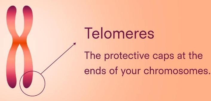
Telomeres are repetitive nucleotide sequences found at the ends of linear chromosomes.
In humans, the telomeric DNA sequence consists primarily of repeated units of:
5′-TTAGGG-3′
These sequences are repeated thousands of times, although the number of repeats varies between chromosomes, individuals, and cell types.
Telomeres are associated with a group of specialized proteins known as the shelterin complex. This protein complex protects chromosome ends and helps prevent them from being mistakenly recognized as damaged DNA.
An important characteristic of telomeres is that they generally become shorter as many human cells undergo repeated divisions. This phenomenon has attracted considerable scientific interest because of its association with cellular ageing, tissue regeneration, cancer, and several age-related disorders.
During DNA replication, DNA polymerase cannot completely replicate the very ends of linear chromosomes. This is known as the end-replication problem.
2. Why Do Telomeres Become Shorter During Cell Division?
The primary reason for telomere shortening is a limitation in the normal DNA replication machinery known as the end-replication problem.
To understand this phenomenon, we first need to examine how DNA replication occurs.
DNA replication takes place during the S phase of the cell cycle. The enzyme DNA polymerase synthesizes new DNA strands using existing DNA strands as templates.
However, DNA polymerase has two important characteristics:
- It can synthesize DNA only in the 5′ to 3′ direction.
- It requires an existing primer with a free 3′-OH group to initiate DNA synthesis.
Because DNA strands are antiparallel, replication occurs differently on the leading and lagging strands.
The leading strand is synthesized continuously, whereas the lagging strand is synthesized discontinuously in short fragments known as Okazaki fragments.
After RNA primers are removed from the lagging strand, the gaps are filled with DNA wherever possible. However, the final RNA primer at the extreme end of the chromosome cannot be replaced with DNA because there is no upstream 3′-OH group available for DNA polymerase to extend from.
Consequently, a small portion of DNA at the chromosome end remains unreplicated.
This results in progressive telomere shortening after successive rounds of DNA replication.
Importantly, telomere shortening is not simply a consequence of DNA polymerase being inaccurate. It is a fundamental limitation associated with replicating the ends of linear DNA molecules.
Other factors, including oxidative stress, inflammation, and DNA damage, can also contribute to telomere attrition.
3. Does this happen in every type of human cell?
| Cell type | Telomere shortening |
| Skin cells | Generally yes |
| Intestinal epithelial cells | Generally yes |
| Blood-forming stem cells | Yes, but telomerase provides some protection |
| Germ cells (sperm and egg precursors) | Telomerase helps maintain telomeres |
| Embryonic cells | High telomerase activity |
| Most mature neurons | Very little cell division, so little division-related shortening |
| Skeletal muscle cells | Mature muscle fibres rarely divide |
4. What is the role of telomerase?
If telomeres shorten during DNA replication, how do certain cells maintain their telomere length?
The answer is an enzyme called telomerase.
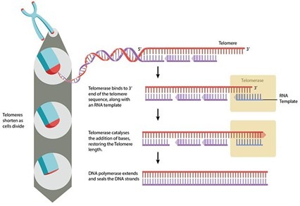
Telomerase is a ribonucleoprotein enzyme complex that adds repetitive DNA sequences to chromosome ends.
It contains two major functional components:
1. TERT (Telomerase Reverse Transcriptase)
TERT is the catalytic protein component of telomerase. It possesses reverse transcriptase activity, allowing it to synthesize DNA using an RNA template.
2. TERC (Telomerase RNA Component)
TERC provides the RNA template that telomerase uses to add new telomeric DNA repeats.
Telomerase binds to the 3′ end of a chromosome and extends the telomeric DNA by adding additional TTAGGG repeats.
Conventional DNA replication machinery can subsequently synthesize the complementary strand.
Through this mechanism, telomerase compensates for the DNA lost during replication.
Telomerase activity is particularly important in:
- Germline cells
- Early embryonic cells
- Certain adult stem cell populations
- Some immune cell populations following activation
- Many cancer cells
However, telomerase does not necessarily maintain telomeres at a constant length in every cell where it is present. The level of activity and the balance between telomere loss and maintenance determine the final outcome.
5. What Happens When Telomeres Become Critically Short?
Telomeres function as protective chromosome caps. However, their protective capacity decreases when they become critically short or dysfunctional.
When this happens, the cell may interpret the chromosome end as damaged DNA.
This can activate DNA damage response pathways involving proteins such as ATM, ATR, p53, and p21.
These pathways can lead to two major cellular outcomes.
A. Cellular senescence
Cellular senescence is a state in which a cell permanently stops dividing while remaining metabolically active.
Senescent cells are not necessarily dead. They can remain present in tissues and release various signaling molecules.
These molecules are collectively known as the senescence-associated secretory phenotype (SASP).
The SASP may include:
- Inflammatory cytokines
- Chemokines
- Growth factors
- Matrix-remodeling enzymes
Senescence has an important protective function because it prevents cells with potentially unstable chromosomes from continuing to proliferate.
However, the accumulation of senescent cells in ageing tissues may contribute to chronic inflammation, impaired tissue regeneration, and age-associated dysfunction.
B. Apoptosis
In some circumstances, severe telomere dysfunction activates programmed cell death, known as apoptosis.
Apoptosis removes damaged or dysfunctional cells and helps maintain tissue health.
The decision between senescence and apoptosis depends on the cell type, the extent of telomere dysfunction, and the molecular signaling pathways activated.
6. What Is the Relationship Between Telomeres and Human Ageing?
Telomere shortening has become an important area of research in the biology of ageing.
As humans grow older, many proliferating tissues show a general tendency toward shorter telomeres.
This is partly associated with repeated cellular divisions throughout life.
However, chronological ageing and telomere shortening are not identical processes.
Not every individual experiences the same rate of telomere shortening. Factors such as genetics, oxidative stress, inflammation, environmental exposures, and lifestyle may influence telomere dynamics.
Furthermore, telomere length varies among different tissues and cell populations within the same individual.
For these reasons, telomere length is considered a potential biomarker of biological ageing, but it is not a perfect or universally reliable measure of an individual’s biological age.
It is also important to recognize that ageing is a complex biological process involving many interacting mechanisms, including:
- Genomic instability
- Epigenetic alterations
- Mitochondrial dysfunction
- Loss of proteostasis
- Cellular senescence
- Stem cell exhaustion
- Chronic inflammation
Telomere shortening is one component of this broader network of ageing mechanisms.
7. The Interesting Connection Between Telomeres and Cancer
There is an important paradox in telomere biology.
On one hand, telomere shortening can protect the body against cancer by limiting the proliferation of potentially abnormal cells.
On the other hand, critically dysfunctional telomeres can promote chromosome instability, which may contribute to cancer development if appropriate cell-cycle checkpoints fail.
Many cancer cells overcome the normal limitations of cellular proliferation by reactivating telomerase.
In fact, telomerase is active in the majority of human cancers.
By maintaining telomere length, cancer cells can continue dividing far beyond the normal proliferative lifespan of many somatic cells.
Some cancers use an alternative mechanism known as alternative lengthening of telomeres (ALT), which maintains telomeres through recombination-based processes rather than conventional telomerase activity.
This is one reason why telomerase and telomere biology have attracted considerable interest in cancer research.
Researchers are investigating whether targeting telomere maintenance mechanisms could provide therapeutic opportunities against cancer.
However, because telomerase is also important in normal stem cells and regenerative tissues, developing safe treatments remains challenging.
8. Can Lifestyle Influence Telomere Length?
Scientific research has investigated whether lifestyle and environmental factors influence telomere maintenance.
Several factors have been associated with differences in telomere length, including:
- Chronic psychological stress
- Smoking
- Obesity
- Chronic inflammation
- Oxidative stress
- Physical activity
- Dietary patterns
For example, smoking and chronic inflammatory conditions have been associated with increased telomere attrition in some studies.
Regular physical activity and healthier lifestyle patterns have also been associated with more favorable telomere-related outcomes in certain populations.
However, these relationships do not necessarily establish direct causation.
There is currently no scientifically established lifestyle intervention that can reliably reverse biological ageing by lengthening telomeres throughout the human body.
Therefore, telomere length should not be viewed as a simple measure that can be controlled through a single dietary supplement or lifestyle modification.
Conclusion: Do Telomeres Shorten During Every Cell Division?
The answer is that telomeres generally shorten during repeated cell divisions in many human somatic cells, but this phenomenon is not universal.
The end-replication problem explains why telomeres progressively shorten in many proliferating cells. However, telomerase can compensate for telomere loss in certain cell populations, including germline cells, early embryonic cells, and some stem cells.
Meanwhile, mature neurons and skeletal muscle fibres generally undergo little or no mitotic division, so they do not experience the usual division-associated telomere shortening.
When telomeres become critically short or dysfunctional, cells may undergo senescence or apoptosis. These processes help prevent the proliferation of genetically unstable cells but may also contribute to age-related tissue dysfunction.
Ultimately, telomeres represent an important link between DNA replication, chromosome stability, cellular lifespan, and human ageing.
Understanding their biology not only helps explain how cells regulate their proliferative capacity but also provides valuable insights into cancer, regenerative medicine, stem cell biology, and age-associated diseases.
The central lesson is that telomere shortening is a regulated and cell-type-dependent biological process, not an inevitable event occurring at the same rate in every cell of the human body.
Key Take-Home Messages
- Telomeres are repetitive DNA sequences that protect the ends of linear chromosomes.
- The end-replication problem is a major reason for telomere shortening during cell division.
- Most proliferating somatic cells have limited telomerase activity and generally experience progressive telomere shortening.
- Telomerase helps maintain telomeres in germline cells, embryonic cells, and certain stem cell populations.
- Mature neurons and skeletal muscle fibres generally do not undergo regular mitotic division.
- Critically short or dysfunctional telomeres can trigger cellular senescence or apoptosis.
- Telomere shortening is associated with ageing, but it is only one component of the complex biology of ageing.
- Many cancer cells reactivate telomerase to maintain their telomeres and sustain uncontrolled proliferation.
Suggested References for Further Reading
- Blackburn, E. H. (1991). Structure and function of telomeres. Nature, 350, 569–573.
- Greider, C. W., & Blackburn, E. H. (1985). Identification of a specific telomere terminal transferase activity in Tetrahymena extracts. Cell, 43(2), 405–413.
- Shay, J. W., & Wright, W. E. (2019). Telomeres and telomerase: Three decades of progress. Nature Reviews Genetics, 20, 299–309.
- López-Otín, C., Blasco, M. A., Partridge, L., Serrano, M., & Kroemer, G. (2013). The hallmarks of aging. Cell, 153(6), 1194–1217.
- Blackburn, E. H., Epel, E. S., & Lin, J. (2015). Human telomere biology: A contributory and interactive factor in aging, disease risks, and protection. Science, 350(6265), 1193–1198.